• 2D
- Tissue Harmonic Imaging (THI)
• Color Doppler
• Power Doppler
• DTI (Doppler Tissue) PW spectral Doppler mode
(for cardiac option)
• PW Doppler
- Duplex Doppler
- Triplex Doppler
• Doppler Tissue Imaging – Color and PW
• CW (Continuous Wave) spectral Doppler
mode (for cardiac option)
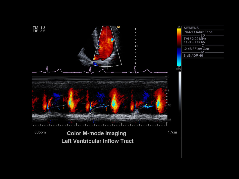
• M-mode and color Doppler M-mode
• MultiHertz™ multiple frequency imaging
• SieScape™ panoramic imaging
• Color SieScape™ panoramic imaging
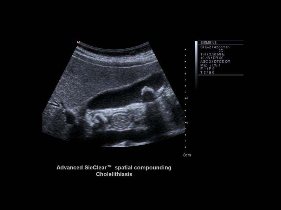
• Advanced SieClear™ spatial compounding
• eSie Touch™ elasticity imaging
• syngo® Velocity Vector Imaging™ technology
• syngo® Arterial Health Package (AHP)*
• syngo® Auto OB Measurements
• Axius™ direct ultrasound research interface
• TEQ™ ultrasound technology for 2D and
spectral Doppler
• SieClear™ multi-view spatial compounding
• 3-Scape™ real-time 3D imaging
• Cadence™ contrast pulse sequencing
technology**
• Clarify™ vascular enhancement technology
• Extend imaging technology
• Advanced fourSight technology
• Gain: -20 to 60 dB in one-decibel increments
• Depth gain compensation: eight DGC controls
• Up to eight user-selectable 2D maps (A – H)
• Up to twelve user-selectable 2D tint maps
(0 – 11)
• Dynamic Tissue Optimization: 4 levels
(Off, 1-3) (for cardiac option)
2D Image Display
• Full screen, dual and seamless dual screen
formats
• Invert (U/D) and transposed (L/R) for all formats
• Image depth up to 30 cm in 0.5 – 1.0 cm
increments (may vary with transducer)
• Sector: selectable field-of-view from 15 to
90 degrees
• Trapezoidal: selectable field of view up to
60 degrees on linear transducers
• Steerable linear: variable steering angles of
2D, color and Doppler modes
• Curved: selectable field of view from 15
to 174 degrees, in one-degree increments,
depending on transducer
• Read/write zoom with image pan
- Available on live, frozen, cine, dual screen
images
- Preserves full image resolution within the
zoom ROI
- Up to 10 X zoom
• On-screen timer (00:00:00)
Color Doppler
• Available on all imaging transducers
• Advanced adaptive processing in color mode
resulting in excellent spatial resolution and
superior flash suppression
• Up to 4 user-selectable transmit frequencies
• Up to 6 user-selectable color velocity maps
(3 velocity; 3 velocity/variance)
• PRF range: 100 to 19,500 Hz
• Up to 512 color samples per color flow
data line
• Gain: -20 to 20 dB in one-decibel increments
• Four wall filter selections (0 – 3)
• Velocity range: 0.004 cm/sec to 450 cm/sec
• Up to 512 2D-mode lines plus 256 color
flow lines
• Up to 5 levels tissue/color priority (0 – 4)
• Up to 4 levels of color smoothing (0 – 3)
• Flow states: Low, General, High for all
applications
• Color persistence levels: Up to 5 levels (0 – 4)
• Resolution/frame rate: 6 levels (0 – 5)
• Color invert
• Color display on/off
Power Doppler Imaging
• Available on all imaging transducers
• Up to 4 user-selectable transmit frequencies
• PRF range: 100 to 19,500 Hz
• Gain: -20 to 20 dB in one-decibel increments
• Four wall filter selections (0 – 3)
• Derived waveform Doppler trace function
analyzes frozen Doppler spectra for mean
and maximum velocity information. Waveform
may be set to trace above baseline, below
baseline or both
• Auto-Doppler trace and calculations performed
in real-time provide auto-calculation and
display of PS, ED, TAMx, TAMn, PI, RI and S/D
Pulsed Wave Doppler Display
• Full screen trace or 2D, 2D/D mode,
simultaneous 2D/C/Doppler and update
• 2D/CW, simultaneous or update
(for cardiac option)
• CW (for cardiac option)
• Four imaging formats: 1/3-2/3, 2/3-1/3, 1/2-1/2,
side by side